Cell structure and organisation
First of all, all organisms are mode of cells. They are like the lego blocks of life. The syllabus wants you to know how to draw a basic animal and plant cell, label its structures, and also explain the functions of each of the structures too.
At a very basic level, please refer to the diagram was below. The plant cell has everything that an animal cell has, plus some added structures which are are in green text. The functions of each of these structures will be discussed further down the page.
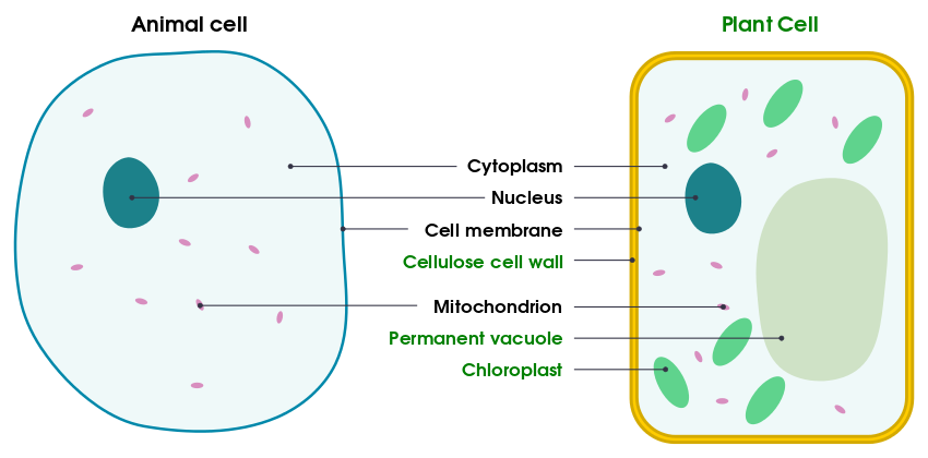
So all cells have a cell membrane which is what allows or disallows certain things entering and exiting the cell. The nucleus contains genetic information (DNA) and the cytoplasm is a jelly-like substance in which everything else in the cell is suspended in. The mitochondrion is the “power house’ of the cell and the reason for this name is due to the fact that respiration occurs here. Plants have some extra structures such as cell walls (to support the cell) and chloroplasts for photosynthesis. You will learn more about these in future topics. Plants also have a permanent vacuole, whereas animal cells have small temporary ones.
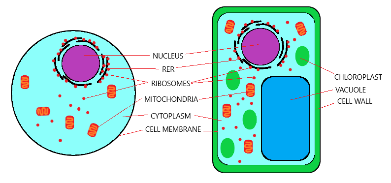
Whilst the above diagram but be sufficient for the core syllabus, the extended course wants you to know two extra structures:
- Rough endoplasmic reticulum (RER)
- Ribosomes
The RER is basically a set of tubular membranes near the nucleus which have ribosomes studded onto it, and the ribosomes are then used for protein synthesis.
These ribosomes can ether be found on the RER (as mentioned before) but it can be found free in the cytoplasm as well. If you are comfortable with the first diagram, take a look at this one!
Structure & function summary
- Cell membrane – Selective control of what goes in and out of the cell
- Nucleus – Carries genetic material (DNA)
- Cytoplasm – Jelly like substance in which chemical reactions take place
- Vacuole – The vacuole has many functions
- Stores/isolates harmful material
- Stores small nutrients
- Maintains water balance
- Structural support for plant cells via turgor pressure
- Rough endoplasmic reticulum – Studded with ribosomes
- Ribosomes – Site of protein synthesis
- Mitochondria – Site of aerobic respiration (cells with high metabolism rates will need lots of these to offer sufficient energy)
- Cell wall – Structural support for plant cells
- Chloroplast – Site of photosynthesis in plant cells
Levels of organisation
There are levels of organisation that you need to be aware of. As we discussed before, the smallest unit of a living thing is a cell. So that’s a good place to start. A group of cells are called tissues, a group of tissues are called organs, and a group of organs are then called organ systems. Take a look here:
- Cell – The smallest structural and functional unit of an organism
- Tissue – Group of cells with similar structures working together to perform a shared function
- Organ – Structure made up of a group of tissues, working together to perform specific functions
- Organ system – Group of organs with related functions, working together to perform body functions
Now some cells have structures that help them with their particular function. There are a couple of these examples that CIE wants you to know:
- Ciliated cells
- Root hair cells
- Xylem vessels
- Palisade cells
- Nerve cells
- Red blood cells
- Sperm and egg cells
Each of the things above will naturally be covered in more detail in other topics in the syllabus and therefore will not be covered here.
Size of specimens
In the lab, a lot of biology is done under a microscope. For example, we can’t exam the cells of a human tissue with our naked eyes right? Therefore the purpose of the microscope is to magnify ourspecimen so that it appears bigger for us to be able to actually see.
Naturally, the CIE syllabus wants you to be able to perform basic equations regarding magnification, the image size (of the specimen) and the actual size (of specimen). Please memorize the following equation:
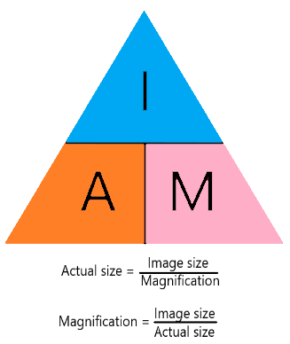
In an exam, they will always give you 2 out of the 3 factors in the equation and tell you to find the missing one. Just apply the formula above and it will be a walk in the park!